

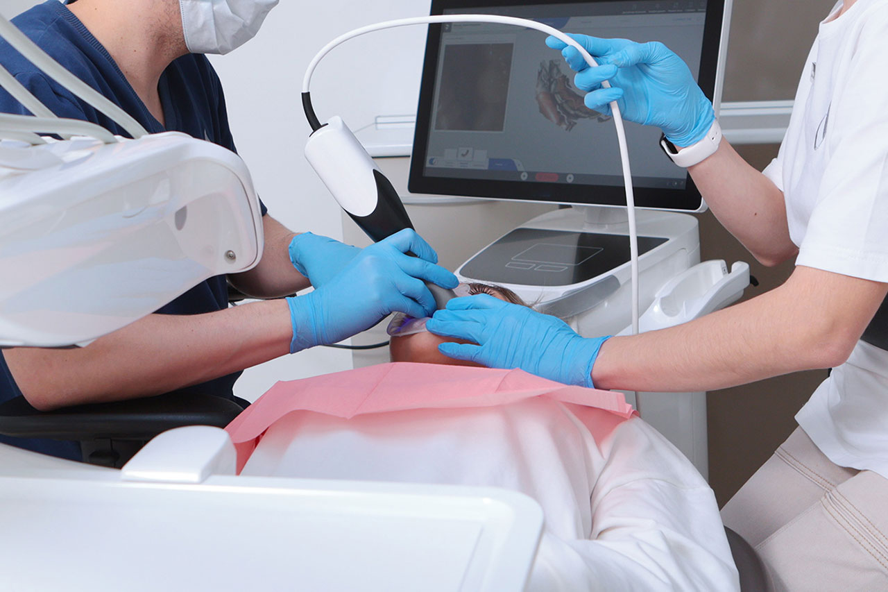
Not every dental problem can be seen in a mirror. A tooth may look healthy on the outside while decay develops between teeth. An infection may begin near a root without causing obvious pain. Changes in the jawbone can also progress before they become noticeable during daily activities. Digital dental X-rays help dentists look beyond visible tooth surfaces to understand what is happening underneath and identify problems that might otherwise remain hidden.
At Dogwood Dental in Marietta, GA, preventive dentistry services include dental examinations and diagnostic imaging when appropriate. Digital dental X-rays are an important part of comprehensive care because they help Dr. Jacobs evaluate areas that cannot be examined by sight alone, supporting earlier diagnosis and more informed treatment decisions.
What Are Digital Dental X-Rays?
Digital dental X-rays are images taken using electronic sensors or digital imaging equipment instead of traditional photographic film. The images show teeth, tooth roots, portions of the jawbone, and surrounding structures.
They allow dentists to evaluate areas that are difficult or impossible to inspect during a standard visual examination.
The process is generally quick. A small sensor or imaging device is positioned to capture the necessary view, and the resulting image can be displayed electronically.
Digital dental X-rays are not intended to replace the dental examination. Instead, they add information that helps the dentist understand the condition of the mouth more completely.
When combined with a review of symptoms, clinical findings, and oral health history, these images can support more accurate decisions about prevention and treatment.
Why Dental Problems Can Develop Without Obvious Symptoms
One of the challenges of dental care is that pain does not always appear when a problem begins.
For example, a cavity between two teeth may grow gradually. Because the area is difficult to see, a patient might not realize anything is wrong until sensitivity develops or a piece of the tooth breaks.
Some infections near tooth roots can also remain relatively quiet for a period of time. Gum disease may cause changes in the bone surrounding teeth before those changes are obvious to the patient.
Digital dental X-rays provide information about these less visible areas. They can help identify developing concerns while a wider range of treatment options may still be available.
Early detection does not mean every finding requires immediate drilling or surgery. Some early changes may be monitored or managed with preventive care, depending on the diagnosis.
What Can Digital Dental X-Rays Reveal?
Different dental images answer different questions. Depending on the type of X-ray, dentists may be able to identify several important concerns.
Cavities Between Teeth
Decay often develops where two teeth touch. These spaces are difficult to examine directly, even with good lighting and dental instruments.
Certain digital dental X-rays can show changes in tooth structure that suggest decay between neighboring teeth. Detecting these areas early may help prevent the need for larger restorations later.
Infection Near Tooth Roots
When the inner tissue of a tooth becomes infected, changes may develop in the bone surrounding the root tip.
Dental imaging can help dentists evaluate these areas when symptoms or clinical findings suggest an infection.
Bone Loss Associated With Gum Disease
Periodontal disease can damage the bone that supports teeth. Digital dental X-rays may reveal changes in bone levels that help the dentist evaluate periodontal health.
These findings are considered alongside gum pocket measurements, bleeding, tooth mobility, and other examination results.
Impacted or Developing Teeth
Some teeth do not emerge into the mouth normally. Wisdom teeth, for example, may remain partially or fully beneath the gums.
Imaging can help show their position and relationship to nearby teeth or structures.
Problems Beneath Existing Dental Work
Crowns, bridges, and fillings can sometimes hide areas where decay or other problems develop.
Digital dental X-rays may help identify changes around restorations that require further evaluation.
Different Types of Dental X-Rays
Dental offices use different types of imaging because each provides information about a particular area.
Bitewing X-Rays
Bitewing images are commonly used to examine the crowns of upper and lower teeth together. They are particularly helpful when checking for cavities between teeth and evaluating some aspects of bone levels.
Periapical X-Rays
These images show the full length of a tooth, including its root and surrounding bone. They may be used when a patient has tooth pain, a suspected infection, or concerns about a particular tooth.
Panoramic X-Rays
A panoramic image provides a broad view of the jaws, teeth, and surrounding structures. It can be useful when evaluating tooth development, wisdom teeth, and certain larger treatment needs.
Three-Dimensional Dental Imaging
Some cases require more detailed imaging. Cone beam computed tomography, often called CBCT, can create three-dimensional views of teeth and jaw structures.
This technology may be considered for certain implant, surgical, or complex diagnostic needs. It is not required for every routine dental examination.
Are Digital Dental X-Rays Safe?
Patients sometimes ask about radiation exposure during dental imaging. This is a reasonable question, particularly for parents and people who have received several medical images.
Digital dental X-rays generally use relatively low radiation doses, and many digital systems require less radiation than older film-based methods for comparable images.
However, dental X-rays still involve ionizing radiation. They should be taken when clinically justified, using appropriate equipment and techniques that keep exposure as low as reasonably achievable while obtaining useful diagnostic information.
Your dentist considers factors such as your age, symptoms, cavity history, gum health, and previous images before recommending new ones.
Inform the dental team if you are pregnant or may be pregnant. Necessary dental imaging can often be performed safely during pregnancy with appropriate clinical judgment, but sharing this information helps guide care.
How Often Should You Have Dental X-Rays?
There is no single imaging schedule that fits every patient.
A person with a history of frequent cavities may require imaging more often than someone with healthy teeth and a low risk of decay.
Children may need images at different intervals as their teeth and jaws develop. Adults with gum disease, dental implants, or a history of complex restorations may also require individualized monitoring.
New patients sometimes need imaging when previous records are not available or when their dental history suggests additional evaluation is necessary.
Before taking new images, the dentist may review existing records to determine whether they already provide the information needed.
The goal is to use digital dental X-rays thoughtfully, obtaining useful information without unnecessary exposure.
Benefits of Digital Dental X-Rays
- Earlier detection: Digital dental X-rays can reveal certain cavities, infections, and bone changes before they cause major symptoms.
- More complete examinations: Imaging provides information about areas that cannot be seen during a visual check.
- Detailed treatment planning: Images help dentists evaluate tooth roots, surrounding bone, and other important structures.
- Efficient image review: Digital images can be displayed quickly and enlarged to discuss findings with patients.
- Convenient record storage: Electronic images can be preserved for comparison during future visits.
- Reduced radiation compared with some older systems: Many digital imaging methods require lower exposure than traditional film techniques.
- Better patient understanding: Patients can often view their images and learn why a particular treatment or monitoring plan is recommended.
How Digital Imaging Supports Preventive Dentistry
Preventive care is most effective when potential problems are recognized before they become severe.
Dental examinations, professional cleanings, fluoride treatments, and good home care form the foundation of prevention. Digital dental X-rays add another layer by helping the dentist identify hidden conditions when imaging is indicated.
For example, an image may reveal a small cavity developing between teeth. If the area is still in an early stage, preventive measures and monitoring may be considered. If the decay has progressed, a filling may be needed before it spreads deeper.
Similarly, imaging may reveal bone changes that encourage a closer periodontal evaluation. Identifying the issue gives the patient an opportunity to address it before additional support is lost.
Digital imaging also supports restorative treatment planning. When replacing a damaged tooth or evaluating an existing crown, the dentist needs a clear understanding of the structures beneath the gums.
These examples show why diagnostic images are helpful tools rather than stand-alone answers.
What to Expect When Getting Dental X-Rays
For many dental images, the process takes only a few minutes. A team member positions a sensor inside the mouth or helps the patient stand or sit correctly for an external image.
You may be asked to bite gently on a holder or remain still while the image is taken.
The procedure itself is generally painless. Some patients experience mild discomfort from holding a sensor in a small mouth or near sensitive tissues. If this occurs, let the dental team know so positioning can be adjusted when possible.
Once the images are available, the dentist can review them as part of the examination and explain any important findings.
Frequently Asked Questions About Digital Dental X-Rays
Do digital dental X-rays hurt?
Taking an X-ray is generally painless. Some patients may feel temporary pressure from the sensor or holder used inside the mouth.
Can dental X-rays detect cavities that are not visible?
Yes. Certain images can help reveal decay between teeth and other changes that may not be visible during a routine examination.
Does every dental checkup require X-rays?
No. Imaging frequency depends on your dental history, symptoms, age, risk factors, and clinical findings.
Are digital dental X-rays useful for dental implant planning?
Yes. Dental imaging helps evaluate jawbone and nearby structures. Some implant cases require three-dimensional imaging for more detailed planning.
Better Information Supports Better Dental Decisions
A healthy mouth depends on more than treating problems that are already painful. Digital dental X-rays help dentists investigate hidden areas, assess oral health changes, and recommend care based on a fuller understanding of each patient’s needs.
When used appropriately with regular examinations and preventive habits, digital imaging can support earlier intervention and more informed treatment planning.
Ready to stay ahead of hidden dental concerns? Contact Dogwood Dental at (770) 926-6886 to book an appointment with Dr. Jacobs at 2421 Shallowford Rd Suite 146, Marietta, GA 30066, and receive personalized preventive dental care for your smile.